You began as a single cell, smaller than a full stop. Today you are made of tens of trillions of cells, and they are not all alike: some are long threads that carry messages, some are tiny red discs that carry oxygen, some wave hairs back and forth all day. How does one cell become so many, and so many different kinds? And how do they work together as one living body?
New cells come from old ones
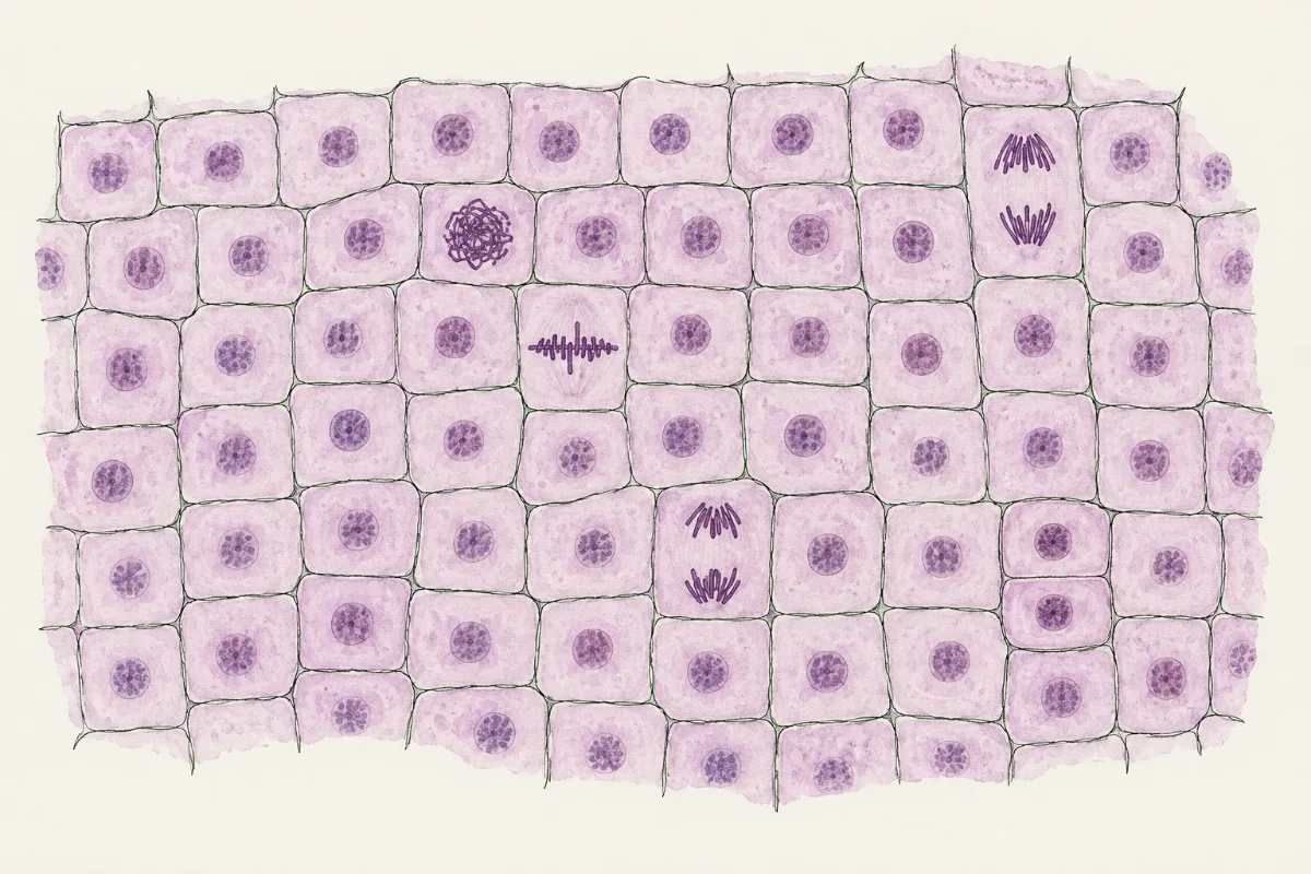
Every new cell is made when an existing cell divides: new cells come only from the division of existing cells. As a body grows, a cell copies its genetic material, then splits in two, so that each new cell gets a full set. In a growing root tip you can catch cells in the act: most look ordinary, but a few have their genetic material packed into dark threads, chromosomes, lined up or pulled apart.
 (enlarge)
Predict first: if every cell comes from a cell that divided, how do the cells of one body end up so different?
Each cell built for its job
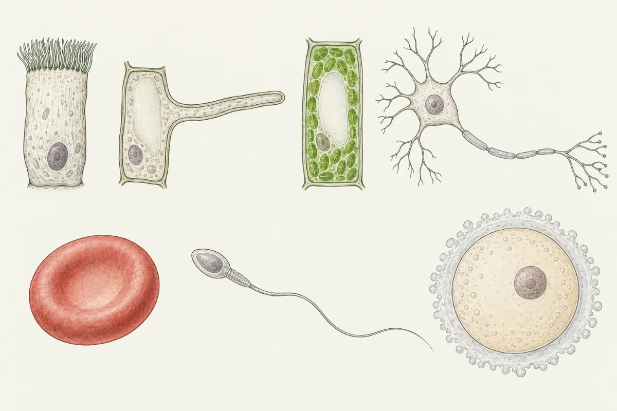
After dividing, many cells change shape and contents to do one job well. This is called becoming specialised. Look at the shapes below and try to guess each job before you read the table.
 (enlarge)
| Cell | Its job | How its structure helps |
|---|---|---|
| Ciliated cell (lining the trachea and bronchi) | moves mucus, with trapped dust and microbes, up and away from the lungs | many tiny hairs, cilia, beat together on one surface |
| Root hair cell | absorbs water and mineral ions from the soil | a long thin extension gives a large surface in contact with the soil water, so more water and ions can enter at once |
| Palisade mesophyll cell (in a leaf) | photosynthesis | packed with chloroplasts, close to the upper surface of the leaf where light falls |
| Neurone (nerve cell) | conducts electrical impulses | a long thin fibre, the axon, carries the impulse a long way |
| Red blood cell | transports oxygen | full of haemoglobin, which carries oxygen; no nucleus, so more room for it; a flattened disc shape gives a large surface for oxygen to pass in and out |
| Sperm cell and egg cell (gametes) | reproduction | the sperm has a tail to swim; the egg is large, with a store of food |
Trap: “A root hair cell is a plant cell, so it has chloroplasts.” It has none: it lives in the dark soil. A cell's structures match its job and where it is.
From cells to a whole organism
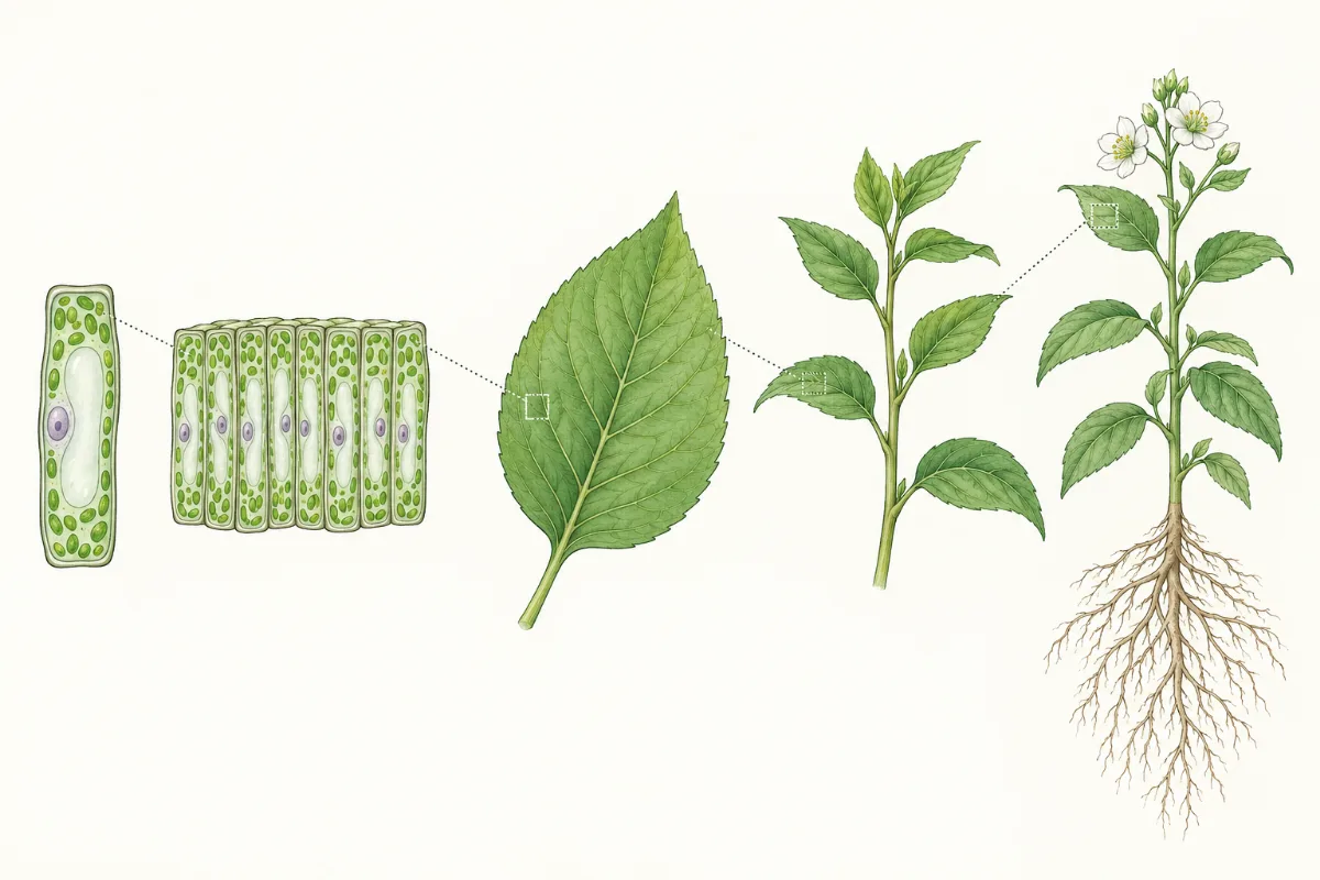
Specialised cells do not work alone. A group of cells with similar structures, working together to do a shared job, is a tissue: the palisade cells of a leaf form palisade tissue. Several tissues working together form an organ, such as the leaf. A group of organs with related jobs, working together, is an organ system, such as the shoot system of a plant or the digestive system of a person. All the organ systems together make the organism. To decide a level, ask what something is made of: similar cells side by side make a tissue; several different tissues make an organ, however small it is.
 (enlarge)
Key idea: cell → tissue → organ → organ system → organism. Each level is made of the level before it, working together.
The same ladder works for you: muscle cells form muscle tissue; muscle, other tissues and a lining form the stomach, an organ; the stomach, intestines, liver and pancreas belong to the digestive system; and all your systems together are you.