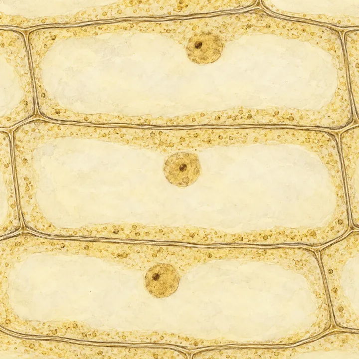
On the snapshot from the microscope, one onion cell stretched across most of the picture: about 164 mm. Yet a real onion cell is smaller than a grain of sugar. So how big is it really? A picture of something tiny is useful only if you know how much it has been enlarged. In this part you measure a cell on a picture and work out its real size, and you learn to write very small sizes in micrometres.
Image size and actual size
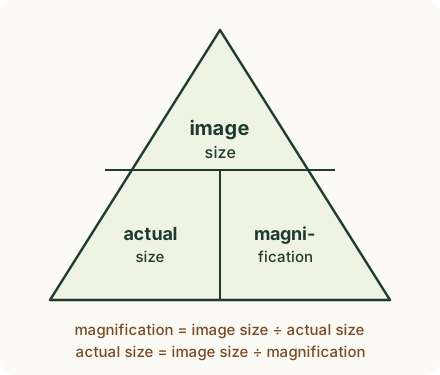
The size of something on a picture or drawing is its image size. Its real size is its actual size. Magnification tells you how many times bigger the image is than the real thing:
Key idea: magnification = image size ÷ actual size
Magnification has no unit: it is written with a ×, for example ×400. Both sizes must be in the same unit before you divide. Rearranging gives actual size = image size ÷ magnification, and image size = magnification × actual size.
 (enlarge)
Predict first: on a picture printed at ×400, is the real cell 400 times bigger or 400 times smaller than its image?
Working out a real size
Your snapshot was printed at ×400, and the ruler gave an image length of about 164 mm (your own reading may differ a little).
actual size = image size ÷ magnification = 164 mm ÷ 400 = 0.41 mm
The real cell is 0.41 mm long: about as long as a printed full stop is wide. The image is always the bigger one (for any magnification above ×1), so the actual size is found by dividing.
(enlarge)Trap: “Multiply the image size by the magnification.” That gives a number far bigger than the picture: 164 mm × 400 = 65 600 mm, more than 65 metres. Check that your answer is smaller than the image.
Magnification can be worked out the other way too. A drawing of a cheek cell is 30 mm across, and the real cell is 0.06 mm across: magnification = 30 mm ÷ 0.06 mm = ×500.
Trap: “The photo is ×400 because the microscope was ×400.” The magnification of a drawing or photo is image size ÷ actual size, not the number on the lenses. Your snapshot happens to be printed at ×400, the same as the microscope; print the same snapshot twice as big and it is ×800. The microscope's ×400 is what you saw in the eyepiece.
Millimetres and micrometres
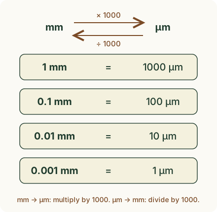
Cells are so small that millimetres give awkward numbers like 0.41 mm or 0.006 mm. Biologists often use micrometres (µm): 1 mm = 1000 µm.
 (enlarge)
So the onion cell is 0.41 mm = 0.41 × 1000 µm = 410 µm long. A cheek cell is about 60 µm across, and a bacterium only 1–5 µm long: about 80 to 400 bacteria, depending on their length, could lie end to end along one onion cell.
To go back from micrometres to millimetres, divide by 1000: 60 µm = 60 ÷ 1000 mm = 0.06 mm. You need this when a question gives the image in millimetres and the real size in micrometres. A drawing of a bacterium is 24 mm long, and the real bacterium is 3 µm long. First 3 µm = 3 ÷ 1000 mm = 0.003 mm; then magnification = 24 mm ÷ 0.003 mm = ×8000.
Trap: “24 mm ÷ 3 µm = ×8.” Before you divide, make sure the image size and the actual size are in the same unit. If the image is in millimetres and the actual size in micrometres, change one of them first.
Scale bars
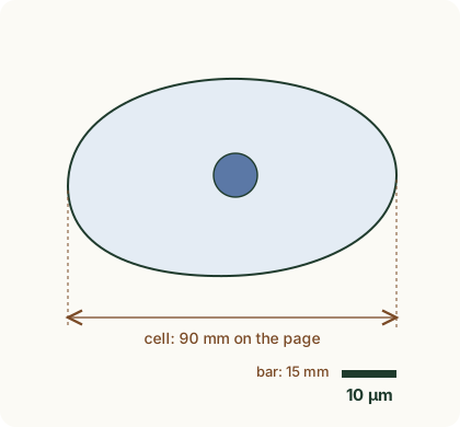
Many photos taken through a microscope, and many drawings, have no × printed on them. Instead they carry a scale bar: a short line labelled with the real length it stands for, such as 10 µm. Measure the bar and the object on the same picture, in millimetres, and work out how many bar lengths fit across the object: each one stands for the real length written on the bar.
 (enlarge)
The cheek cell above is 90 mm across on the page, and the 10 µm bar is 15 mm long. The cell is 90 ÷ 15 = 6 bar lengths wide, so it is really 6 × 10 µm = 60 µm across. You can also find the magnification from the bar: 10 µm = 0.01 mm, so magnification = 15 mm ÷ 0.01 mm = ×1500, and actual size = 90 mm ÷ 1500 = 0.06 mm = 60 µm.
Key idea: actual size = image size ÷ magnification, with both sizes in the same unit. 1 mm = 1000 µm. A scale bar gives the real length of one line on the picture; count how many fit across the object.