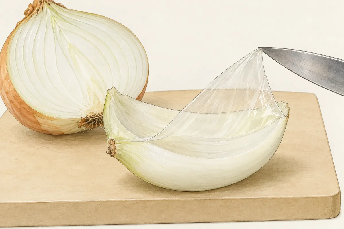
Peel the thin skin from the inside of an onion scale and hold it up to the light. It is as clear as cling film, and you can read through it. Yet under a microscope this "film" turns out to be a wall of tiny boxes, packed side by side like bricks. Your own body is built the same way, from units far too small to see. So what is inside one of those boxes, and is a piece of onion built like a piece of you? In this part you prepare a slide, focus a microscope and compare plant and animal cells.
 (enlarge)
The light microscope
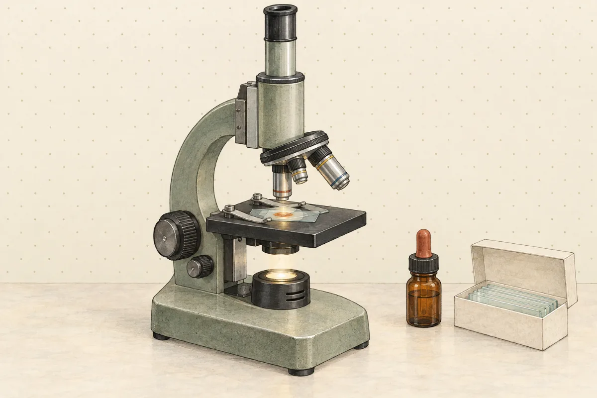
A light microscope shines light up through a thin specimen and magnifies the image with two lenses: the eyepiece you look through, and an objective close to the specimen. Most school microscopes have three objectives on a turning nosepiece. With a ×10 eyepiece, the ×4, ×10 and ×40 objectives give a total magnification of ×40, ×100 and ×400.
 (enlarge)
The specimen must be thin, so that light can pass through it. It lies in a drop of liquid on a glass slide, under a thin square of glass called a coverslip. Lower the coverslip at an angle, from one edge, so that it pushes the air out in front of it. Dropped flat, it traps air bubbles, which look like round black-edged blobs and hide the cells. A stain such as iodine solution colours some parts of the cells so that they are easier to see.
Two knobs focus the image by moving the stage up or down. The large coarse focus knob moves it a long way; the small fine focus knob moves it a tiny distance. Start with the lowest power and the coarse knob. At high power use only the fine knob: the ×40 objective almost touches the slide, and a turn of the coarse knob could crack the slide and scratch the lens.
Predict first. Will you see anything green inside the onion cells?
Did your onion cells have anything green in them? Look again at the ×400 view before you read on.
What you saw: onion and cheek cells
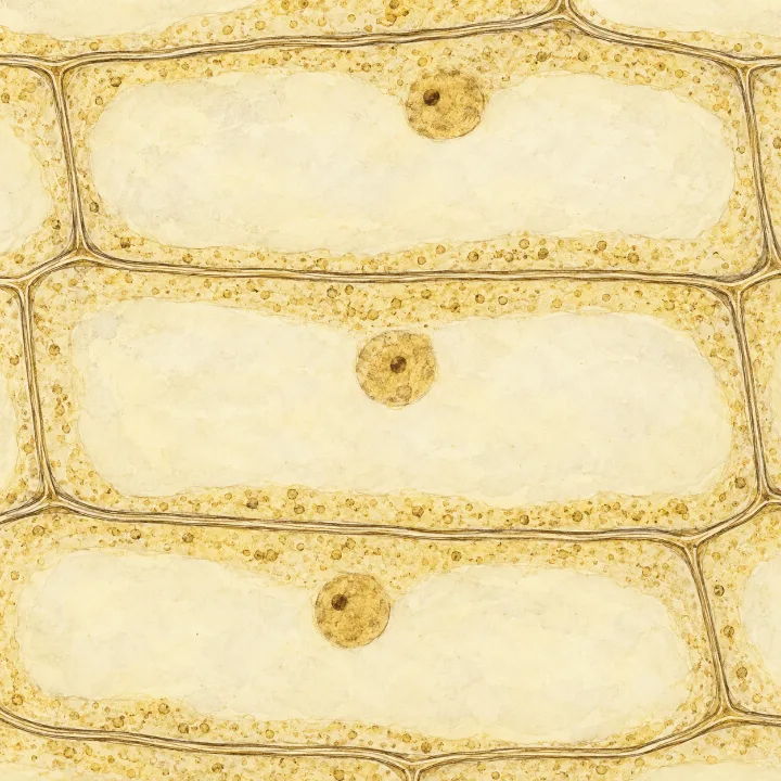
Each onion cell is outlined by its cell wall, a firm layer that keeps the cell's box shape. Inside, a thin layer of cytoplasm lines the wall, and a darker round body, the nucleus, sits in it, often to one side. Most of the cell is filled by a large clear space, the vacuole, full of a watery liquid called cell sap.
 (enlarge)
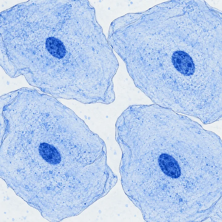
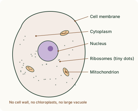
Cheek cells look quite different. They are flat and irregular, and they sit loosely, with gaps between them. Each has a nucleus and cytoplasm, but no cell wall and no large vacuole. The outline you see round each cheek cell is its cell membrane, the very thin layer that every cell has around its cytoplasm. A plant cell has one too, pressed against the inside of its wall, so in an onion cell you see the two as one line.
 (enlarge)
Trap: “It's a plant, so its cells must be green.” Not every plant cell is green. Onion skin is part of a storage leaf inside the bulb, where no light reaches, so its cells have no chloroplasts.
The green cells
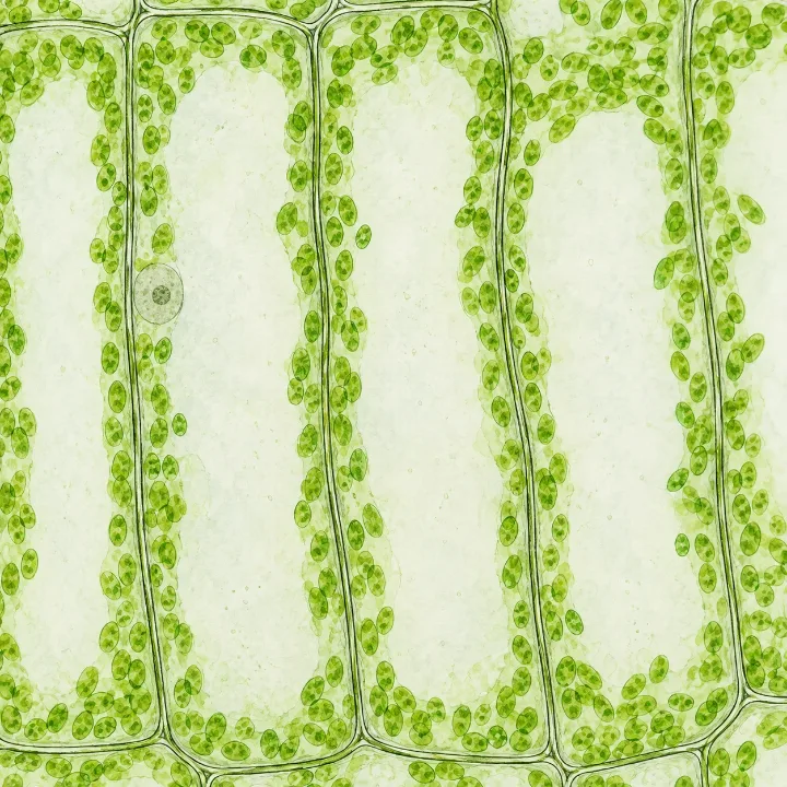
The pondweed leaf answers the green question. Its cells have walls and vacuoles like the onion cells, but they are full of small green bodies lying along the walls: chloroplasts. Chloroplasts contain the green pigment chlorophyll and are where photosynthesis happens: the plant uses the energy of light to make sugar.
 (enlarge)
Inside plant and animal cells
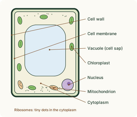
A light microscope shows walls, nuclei, cytoplasm, vacuoles and chloroplasts. With an electron microscope, which can magnify far more, biologists also see much smaller structures. Two of them are in both plant and animal cells: mitochondria, where aerobic respiration (a reaction that uses oxygen) releases energy from food, and ribosomes, tiny grains where proteins are made.
 (enlarge)
 (enlarge)
| Structure | What it does | Plant cell | Animal cell |
|---|---|---|---|
| Cell wall | made of cellulose; keeps the cell's shape and stops it bursting | yes | no |
| Cell membrane | controls what enters and leaves the cell | yes | yes |
| Nucleus | contains the genetic material (DNA) that controls the cell | yes | yes |
| Cytoplasm | jelly-like; most of the cell's chemical reactions happen here | yes | yes |
| Chloroplasts | photosynthesis (only in cells of green parts) | some cells | no |
| Large (permanent) vacuole | stores cell sap; keeps the cell firm; lasts for the cell's life | yes | no (only small ones, if any) |
| Mitochondria | aerobic respiration: release energy | yes | yes |
| Ribosomes | make proteins | yes | yes |
Key idea: Plant and animal cells both have a cell membrane, cytoplasm, a nucleus, mitochondria and ribosomes. Only plant cells have a cellulose cell wall, a large permanent vacuole and, in green parts, chloroplasts.
Trap: “The cell wall controls what enters the cell.” Water and dissolved substances pass through the wall easily; the cell membrane is the layer that controls entry and exit.
A cell without a nucleus
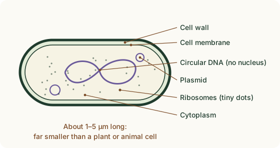
Bacteria are single cells, and much smaller than plant or animal cells: most are 1 to 5 µm long (a micrometre, µm, is a thousandth of a millimetre). They have a cell wall, a cell membrane, cytoplasm and ribosomes, but no nucleus. Their DNA is a single loop, the circular DNA, lying in the cytoplasm, and many bacteria also carry extra small rings of DNA called plasmids. The circular DNA carries the genes, the instructions that control the cell. A plasmid carries only a few extra genes, for example one that lets the bacterium survive an antibiotic.
The other parts do the same jobs as in a plant cell: the wall (not made of cellulose) keeps the cell's shape and stops it bursting, the membrane controls what enters and leaves, and the ribosomes make proteins. With a school microscope at ×400, bacteria look at most like tiny dots or rods: their inner parts can only be seen with an electron microscope.
 (enlarge)
Key idea: To identify a cell in a drawing or photo, first look for a nucleus. A tiny cell with a wall, no nucleus and its DNA in a loose loop: a bacterium. A nucleus and a firm wall outside the membrane, often with a large vacuole or green chloroplasts: a plant cell. A nucleus and only a membrane round the cytoplasm: an animal cell.